
Phantom Limb Study Rewires our Understanding of the Brain


By Science Correspondent
In a first-of-its-kind study, researchers found that the brain’s control center for a lost appendage can persist long after surgical amputation — a finding that challenges long-held theories about the brain’s ability to reorganize itself, also known as plasticity.
Scientists from the National Institutes of Health (NIH) and University College London examined brain activity before and after arm amputations and found that losing a limb does not trigger a large-scale cerebral overhaul. Published in Nature Neuroscience, the study offers new insight into phantom limb syndrome and could help guide the development of neuroprosthetics and pain treatments.
The team took advantage of a rare opportunity: MRI scans of three participants in the months before planned amputations, followed by scans up to five years after.
“It’s not often you get the chance to conduct a study like this one, so we wanted to be exceedingly thorough,” said co-author Chris Baker, Ph.D., of NIH’s National Institute of Mental Health (NIMH). “We approached our data from a variety of angles and all of our results tell a consistent story.”
The brain’s outermost layer, the cortex, assigns different regions to different body parts. Traditionally, neuroscientists believed that when a body part is lost, neighboring brain areas would “remap” themselves onto the vacant region.
“For many decades, cortical remapping as a response to amputation has been a literal textbook example of brain plasticity,” Baker said.
However, phantom limb syndrome — where individuals feel vivid, often painful sensations in a missing limb — has long complicated this theory. Baker and his team saw this as a clue: perhaps the brain retains its map of the lost limb.
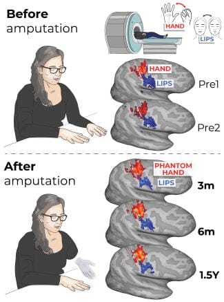
After years of searching, they identified three individuals already scheduled for arm amputation. The team conducted functional MRI scans twice before surgery, mapping brain activity triggered by tapping individual fingers.
They followed up with three post-surgery scans as participants tried to perform the same tasks using their phantom limbs.
They compared brain activity across time points and found little to no change.
Baker explained that without knowing when the data was collected, they likely wouldn't have been able to tell the difference between brain maps.
A machine learning algorithm — trained on pre-amputation data — could still accurately identify which phantom finger was being “moved” after surgery. Circuits tied to other body parts, such as the lips or feet, did not invade the area formerly used by the amputated limb.
Comparisons with able-bodied controls and other studies reinforced the finding: the brain’s representation of the missing limb stays intact.
These results could help explain how phantom limb syndrome occurs and suggest a need to revisit treatments that assume cortical reorganization happens after amputation.
According to lead author Hunter Schone, Ph.D., who conducted the research as a graduate student at NIH, the findings also offer important insights for brain-computer interface technologies.
“This study is a powerful reminder that even after limb loss, the brain holds onto the body, almost like it’s waiting to reconnect in some new way,” said Schone.
“Now, rapidly developing brain-computer interface technologies can operate under the assumption that the brain's body map remains consistent over time. This allows us to move into the next frontier: accessing finer details of the hand map, like distinguishing the tip of the finger from the base, and restoring the rich, qualitative aspects of sensation, such as texture, shape and temperature.”